Sliding filament theory coloring answers
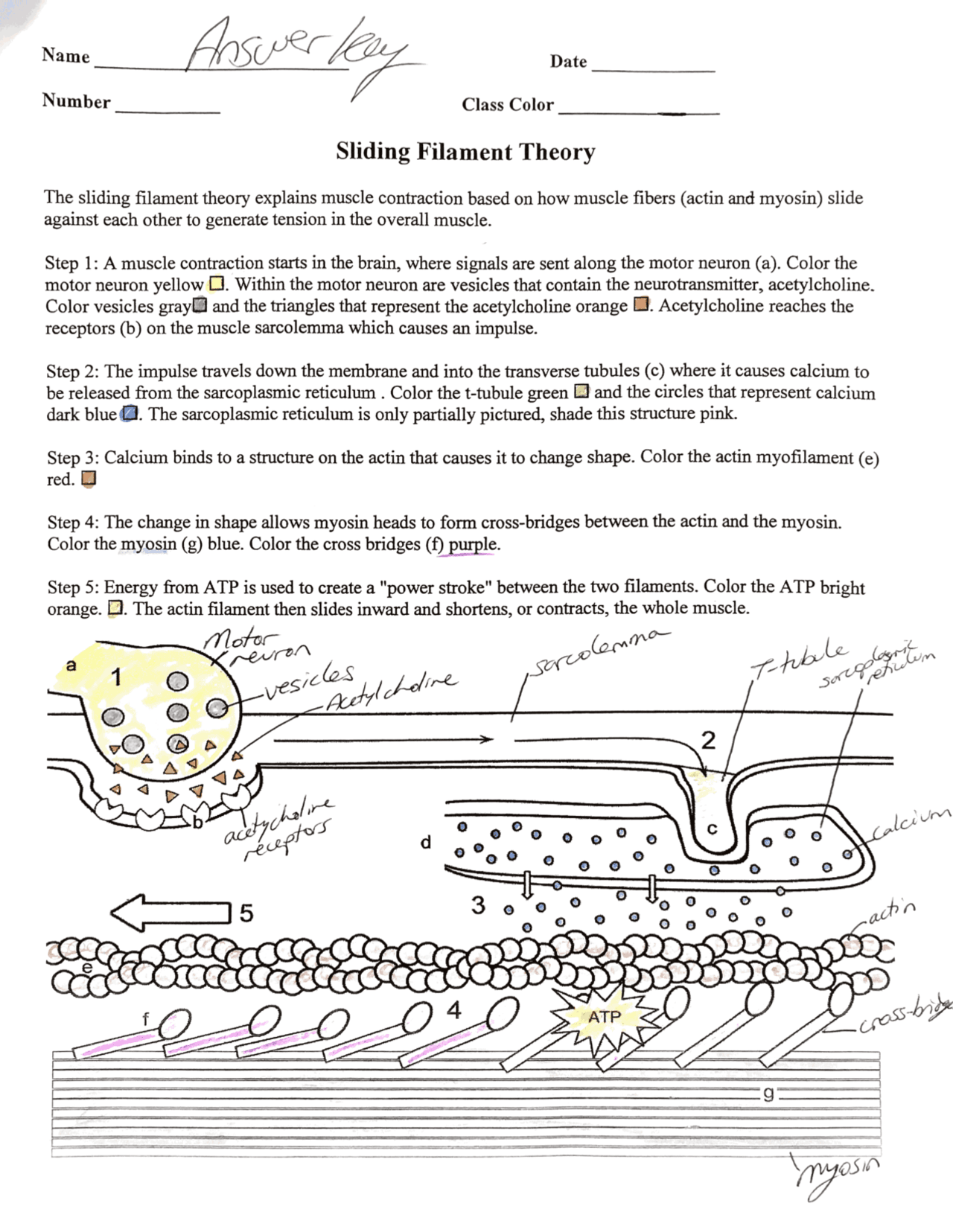
The sliding filament theory explains muscle contraction based on how muscle fibers actin and myosin slide against each other to generate tension in the overall muscle.
Sarcomeres are composed of thick and thin filaments. The thin filament is composed of polymerized actin, while the thick filament is composed of myosin. Titin is a protein that spans the full range of the sarcomere, and is involved in stability and elasticity in the muscle. Collagen is not a primary component of sarcomeres. The protein complex formed is classically named actomyosin and helps facilitate the "stroke" part of muscle contraction. The sliding filament theory describes the mechanism that allows muscles to contract.
Sliding filament theory coloring answers
Each skeletal muscle is an organ that consists of various integrated tissues. These tissues include the skeletal muscle fibers, blood vessels, nerve fibers, and connective tissue. Each skeletal muscle has three layers of connective tissue called mysia that enclose it, provide structure to the muscle, and compartmentalize the muscle fibers within the muscle Figure Each muscle is wrapped in a sheath of dense, irregular connective tissue called the epimysium , which allows a muscle to contract and move powerfully while maintaining its structural integrity. The epimysium also separates muscle from other tissues and organs in the area, allowing the muscle to move independently. Inside each skeletal muscle, muscle fibers are organized into bundles, called fascicles , surrounded by a middle layer of connective tissue called the perimysium. This fascicular organization is common in muscles of the limbs; it allows the nervous system to trigger a specific movement of a muscle by activating a subset of muscle fibers within a fascicle of the muscle. Inside each fascicle, each muscle fiber is encased in a thin connective tissue layer of collagen and reticular fibers called the endomysium. The endomysium surrounds the extracellular matrix of the cells and plays a role in transferring force produced by the muscle fibers to the tendons. In skeletal muscles that work with tendons to pull on bones, the collagen in the three connective tissue layers intertwines with the collagen of a tendon. At the other end of the tendon, it fuses with the periosteum coating the bone. The tension created by contraction of the muscle fibers is then transferred though the connective tissue layers, to the tendon, and then to the periosteum to pull on the bone for movement of the skeleton.
It is the shortening of these individual sarcomeres that lead to the contraction of individual skeletal muscle fibers and ultimately the whole muscle.
This worksheet lists the steps involved in the sliding filament model of muscle contraction and includes a coloring page of the model. Students color and answer questions. This download has two versions of the student worksheet 2 pages vs 1 pages and the answer key to the questions. Log In Join. View Wish List View Cart.
The sliding filament theory explains muscle contraction based on how muscle fibers actin and myosin slide against each other to generate tension in the overall muscle. Step 1 : A muscle contraction starts in the brain, where a signal is sent to the motor neuron a. The combination of the motor neuron and the skeletal muscle fibers make up a motor unit. Color the motor neuron a yellow. Vesicles that contain the neurotransmitter, acetylcholine. Color vesicles b gray.
Sliding filament theory coloring answers
This worksheet lists the steps involved in the sliding filament model of muscle contraction and includes a coloring page of the model. Students color and answer questions. This download has two versions of the student worksheet 2 pages vs 1 pages and the answer key to the questions. Log In Join. View Wish List View Cart. Middle school. High school.
Emma starletto vk
Color the triangles that represent the acetylcholine c orange. External Website Watch this video to learn more about macro- and microstructures of skeletal muscles. Color the ATP orange. Sarcomeres are composed of thick and thin filaments. Classroom community. Total Pages. Kindergarten math. IXL family of brands. Items are fill in the blank, short answer and open response. High school social studies. Top Subjects. By topic. Rosetta Stone. Color vesicles b gray.
Sliding Filament Graphic - shows how actin and myosin interact, labeled in stages. Includes questions to answer.
These items are sold separately in my store but I'm offering the complete lesson at a discounted price. These regions represent areas where the filaments do not overlap, and as filament overlap increases during contraction these regions of no overlap decrease. Acetylcholine reaches the receptors b on the muscle sarcolemma which causes an impulse. Clip art. Speech therapy. The actin and myosin filaments slide past one another. Study Guides. Social emotional. Description optional. Where is calcium released from? Interactive Link Questions Watch this video to learn more about macro- and microstructures of skeletal muscles.


This rather good phrase is necessary just by the way
You are not right. I can defend the position. Write to me in PM, we will communicate.
Excuse, that I interrupt you, but, in my opinion, this theme is not so actual.