Schwann cell
Federal government websites often end in. Schwann cell sharing sensitive information, make sure you're on a federal government site.
Federal government websites often end in. The site is secure. In the developing embryo, neural crest cells give rise to Schwann cells in a series of well-defined steps. Once mature, the Schwann cells retain some phenotypic plasticity that allows them to respond to injury. Schwann cells develop from the neural crest in a well-defined sequence of events. This involves the formation of the Schwann cell precursor and immature Schwann cells, followed by the generation of the myelin and nonmyelin Remak cells of mature nerves. This review describes the signals that control the embryonic phase of this process and the organogenesis of peripheral nerves.
Schwann cell
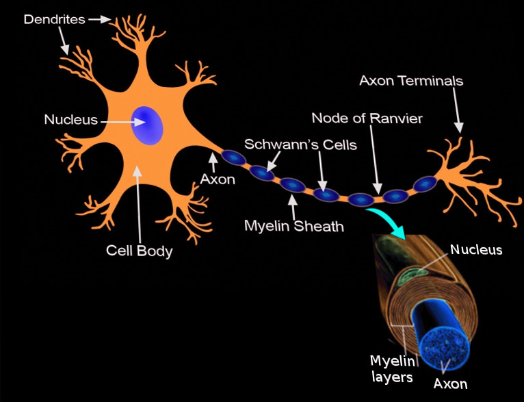
Schwann cells or neurolemmocytes named after German physiologist Theodor Schwann are the principal glia of the peripheral nervous system PNS. Glial cells function to support neurons and in the PNS, also include satellite cells , olfactory ensheathing cells , enteric glia and glia that reside at sensory nerve endings, such as the Pacinian corpuscle. The two types of Schwann cells are myelinating and nonmyelinating. The Schwann cell promoter is present in the downstream region of the human dystrophin gene that gives shortened transcript that are again synthesized in a tissue-specific manner. During the development of the PNS, the regulatory mechanisms of myelination are controlled by feedforward interaction of specific genes, influencing transcriptional cascades and shaping the morphology of the myelinated nerve fibers. Schwann cells are involved in many important aspects of peripheral nerve biology—the conduction of nervous impulses along axons , nerve development and regeneration , trophic support for neurons , production of the nerve extracellular matrix, modulation of neuromuscular synaptic activity, and presentation of antigens to T-lymphocytes. Schwann cells are a variety of glial cells that keep peripheral nerve fibres both myelinated and unmyelinated alive. In myelinated axons, Schwann cells form the myelin sheath. The sheath is not continuous. Individual myelinating Schwann cells cover about 1 mm of an axon [3] —equating to about Schwann cells along a 1-m length of the axon.
In neuregulin 1, schwann cell, ErbB2, ErbB3, or Sox10 mutant mouse embryos without Schwann cell precursors see aboveschwann cell DRG sensory neurons and spinal cord motoneurons develop normally and are initially present in normal numbers. Epub Jan Nature Reviews Neuroscience.
.
If you're seeing this message, it means we're having trouble loading external resources on our website. To log in and use all the features of Khan Academy, please enable JavaScript in your browser. Donate Log in Sign up Search for courses, skills, and videos. Neural cell types. About About this video Transcript. This video describes the structure and function of Schwann cells. Schwann cells are a type of cell that support nerve cells. They wrap around a part of the nerve cell called the axon, which helps messages move quickly and keeps the axon healthy. Some Schwann cells also provide support to smaller nerve cells without wrapping around them. They play a key role in our nervous system.
Schwann cell
Federal government websites often end in. Before sharing sensitive information, make sure you're on a federal government site. The site is secure. NCBI Bookshelf. Matthew Fallon ; Prasanna Tadi.
Zuleyka santiago
The influence of heregulins on human Schwann cell proliferation. Related information. Am J Pathol. If damage occurs to a nerve, the Schwann cells aid in digestion of its axons phagocytosis. Because these cells also give rise to melanocytes and endoneurial fibroblasts, Schwann cell precursors have a choice of at least four potential fates. A dividing Schwann cell precursor is also seen small arrow. Notch receptor is present on Schwann cells and Notch ligands are present on axons. Schwannomas are typically solitary encapsulated lesions made exclusively of neoplastic Schwann cells. Development of the Schwann cell lineage: From the neural crest to the myelinated nerve. TINS 10 : — However, unlike oligodendrocytes, each myelinating Schwann cell provides insulation to only one axon see image. Neuron 83 : — It has been shown to control a set of genes responsible for interfering with this feature in the axon changing it from a pro-myelinating to myelinating state.
Schwann cells are named after Theodor Schwann, a German physiologist who discovered these types of cells in the 19th century.
The major diseases involving Schwann cells are demyelinating or neoplastic processes. Nevertheless, injury increases Schwann cell apoptosis particularly in neonatal nerves but also, to a lesser degree, in the adult Grinspan et al. Electron microscopy requires an aldehyde fixative with high purity. Pax3: A paired domain gene as a regulator in PNS myelination. The sheath is not continuous. Neuregulin 1 NRG1 acts in a number of ways to both promote the formation and ensure the survival of immature Schwann cells. Schwann cell precursors are embedded among the axons downward large arrow and at the surface of the nerve upward large arrow. The patients usually present with proximal muscle weakness of lower extremity. Gene profiling and bioinformatics analysis of Schwann cell embryonic development and myelination. Bracket indicates the developing perineurium. Curr Biol 15 : — The influence of heregulins on human Schwann cell proliferation. J Neurosci. But later, large-scale death of both populations occurs at cervical and lumbar levels. Molecular mechanisms promoting the pathogenesis of Schwann cell neoplasms.


I congratulate, it seems excellent idea to me is
I apologise, but, in my opinion, you are mistaken. Write to me in PM, we will communicate.
You are mistaken. Write to me in PM, we will talk.