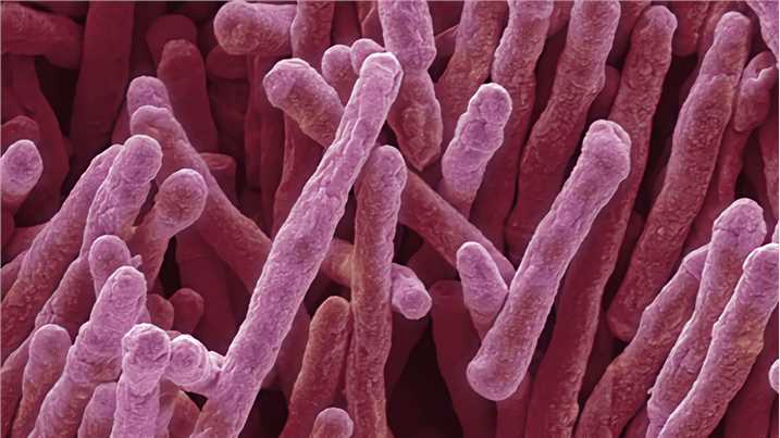
Mycobacterium smegmatis
A series of structome analyses, that is, quantitative and three-dimensional structural analysis of a whole cell at the electron microscopic level, have already been achieved individually in Exophiala dermatitidis, Saccharomyces cerevisiae, Mycobacterium tuberculosisMyojin spiral bacteria, and Escherichia coli, mycobacterium smegmatis.
The genus Mycobacterium contains several slow-growing human pathogens, including Mycobacterium tuberculosis, Mycobacterium leprae, and Mycobacterium avium. Mycobacterium smegmatis is a nonpathogenic and fast growing species within this genus. In , a mutant of M. Classical bacterial models, such as Escherichia coli, were inadequate for mycobacteria research because they have low genetic conservation, different physiology, and lack the novel envelope structure that distinguishes the Mycobacterium genus. By contrast, M.
Mycobacterium smegmatis
Mycobacterium smegmatis is an acid-fast bacterial species in the phylum Actinomycetota and the genus Mycobacterium. It is 3. It was first reported in November by Lustgarten, who found a bacillus with the staining appearance of tubercle bacilli in syphilitic chancres. Subsequent to this, Alvarez and Tavel found organisms similar to that described by Lustgarten also in normal genital secretions smegma. This organism was later named M. Some species of the genus Mycobacterium have recently been renamed to Mycolicibacterium , so that M. Essentially, the bacteria form a single-layered sheet and are able to move slowly together without the use of any extracellular structures, like flagella or pili. For example, this sliding ability is correlated with the presence of glycopeptidolipids GLPs on the outermost part of the cell wall. GLPs are amphiphilic molecules that could potentially decrease surface interactions or create a conditioning film that allows movement. Although the exact role of GLPs in sliding is not known, without them M. Mycobacterium smegmatis is useful for the research analysis of other Mycobacteria species in laboratory experiments.
Singh, A. Then, cells with older growth poles elongate faster than cells with younger growth poles.
.
Thank you for visiting nature. You are using a browser version with limited support for CSS. To obtain the best experience, we recommend you use a more up to date browser or turn off compatibility mode in Internet Explorer. In the meantime, to ensure continued support, we are displaying the site without styles and JavaScript. Two genes, pafB and pafC , are organized in an operon with the Pup-ligase gene pafA , which is part of the Pup-proteasome system PPS present in mycobacteria and other actinobacteria. To characterize their function, we generated a pafBC deletion in Mycobacterium smegmatis Msm. Proteome analysis of the mutant strain revealed decreased cellular levels of various proteins involved in DNA damage repair, including recombinase A RecA. PafB and PafC feature winged helix-turn-helix DNA binding motifs and we demonstrate that together they form a stable heterodimer in vitro , implying a function as a heterodimeric transcriptional regulator.
Mycobacterium smegmatis
Federal government websites often end in. The site is secure. The genus Mycobacterium contains several slow-growing human pathogens, including Mycobacterium tuberculosis , Mycobacterium leprae , and Mycobacterium avium. Mycobacterium smegmatis is a nonpathogenic and fast growing species within this genus. In , a mutant of M.
El segundo aire película completa en español latino online
However, average cytoplasmic ribosome density of M. As mentioned in our previous paper, there is a close correlation between ribosome density and doubling time min Yamada et al. In other projects. Nucleic Acids Res. Methods 9, — These findings can be applied to the Snm secretion system of M. Table 1. HY and MY performed experiments. These effector proteins are important virulence factors, which allow the pathogen to survive inside of the host. Liu et al. Tsaloglou, M. In comparison of cell diameter, both average OM 0.
Thank you for visiting nature. You are using a browser version with limited support for CSS.
Papagiannakis, A. Our data confirm these reports. Goins, C. Gene expression profiling of the TRIM protein family reveals potential biomarkers for indicating tuberculosis status. Authors used M. It is then surprising that there was only a small difference in average cytoplasmic ribosome density between M. In this study, M. These values are approximately twice of those of M. Furthermore, average whole cell diameter of M. Bacteriological Reviews. Mortuza, R. Phylogenomics and comparative genomic studies robustly support division of the genus Mycobacterium into an emended genus Mycobacterium and four novel genera. The volume of the periplasm was calculated by subtracting the cytoplasmic volume from the whole cell volume.


I advise to you to try to look in google.com
Excuse, that I can not participate now in discussion - it is very occupied. I will return - I will necessarily express the opinion on this question.
I apologise, but, in my opinion, you are not right. I am assured. Let's discuss it.