Molar pregnancy radiology
At the time the article was last revised Wedyan Yousef Alrasheed had no financial relationships to ineligible companies to disclose.
Federal government websites often end in. The site is secure. Ectopic molar pregnancy is extremely rare, and preoperative diagnosis is difficult. Our literature search found only one report of molar pregnancy diagnosed preoperatively. Moreover, there is no English literature depicting magnetic resonance image MRI findings of ectopic molar pregnancy. We report a case of ectopic molar pregnancy preoperatively diagnosed using MRI. All patients underwent surgical resection or dilatation and curettage.
Molar pregnancy radiology
At the time the article was last revised Ammar Ashraf had no financial relationships to ineligible companies to disclose. A complete hydatidiform mole CHM is a type of molar pregnancy and falls at the benign end of the spectrum of gestational trophoblastic disease. Complete moles are characterized by the absence of a fetus or fetal parts i. There is a non-invasive, diffuse swelling of chorionic villi. Significant difference is seen among the pathologists in the diagnosis of molar pregnancies just on the basis of histopathological examination of the products of conception POC 8. The p57KIP2 gene is paternally imprinted and expressed from the maternal allele 8,9. Polymer-based immunohistochemistry IHC with p57, shows absent staining in the complete mole CM and positive staining in the hydropic abortus HA and partial mole PM 8,9. This IHC staining is a useful and inexpensive tool which can help in distinguishing complete mole from its mimics and can avoid DNA analysis 8,9. All the chromosomes are derived from the sperm, suggesting fertilisation of a single egg that has lost its chromosomes. Serum beta HCG levels are markedly elevated, out of proportion to the pregnancy. CT evaluation is not usually performed due to its low resolution for the uterine assessment. CT may show an enlarged uterus with areas of low attenuation, or hypoattenuating foci surrounded by highly enhanced areas in the myometrium. MRI may demonstrate a heterogeneous mass with cystic spaces distending the uterine cavity. Fetal parts are notably absent. Uterine zonal anatomy is often distorted although a hypointense irregular myometrial boundary may be seen 3.
Because molar ectopic pregnancy was suspected and her vital signs were stable, MRI was performed. Nucci, Esther Oliva.
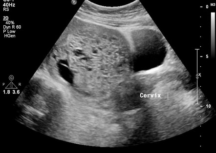
Federal government websites often end in. The site is secure. Ultrasound of a molar pregnancy with long axis view and short axis view. Click here to view. A 32 year-old female presented to the emergency department ED with complaints of mild vaginal spotting accompanied by uterine cramping.
Molar pregnancy, part of the Gestational Trophoblastic Disease spectrum, presents as grape-like placental tissue, markedly elevated hCG levels, the absence of a viable foetus, and a characteristic snowstorm appearance on US due to the presence of numerous small vesicles within the uterus. A molar pregnancy, also known as a hydatidiform mole, is an abnormal form of pregnancy where a fertilised egg fails to develop into a viable foetus and instead grows into a mass of abnormal tissue in the uterus. This condition is part of the spectrum of Gestational Trophoblastic Disease GTD , characterised by abnormal proliferation of placental trophoblasts. Molar pregnancies occur due to anomalous fertilisation events. In a complete mole, an enucleated empty egg gets fertilised by a sperm, which then duplicates its chromosomes, leading to a diploid set, all paternal in origin 46, XX karyotype. Partial moles arise from an egg fertilised by two sperms, resulting in a triploid set of chromosomes, two-thirds of which are paternal 69, XXX or 69, XXY karyotypes. Molar pregnancies are not typically graded or staged but are classified as either complete or partial. Diagnosis is established on the basis of characteristic clinical, sonographic, and biochemical findings.
Molar pregnancy radiology
During a transvaginal ultrasound, you lie on an exam table while a doctor or a medical technician puts a wandlike device, known as a transducer, into the vagina. Sound waves from the transducer create images of the uterus, ovaries and fallopian tubes. A health care provider who suspects a molar pregnancy is likely to order blood tests and an ultrasound.
Leaguesecretary
Journal of Minimally Invasive Gynecology. Ikuma et al. Gillespie et al. Fallopian tube invasive molar disease. At the time the article was created The Radswiki had no recorded disclosures. Dumitrescu and A. Associated Data Supplementary Materials Video. Moreover, there is no English literature depicting magnetic resonance image MRI findings of ectopic molar pregnancy. Journal of the Turkish German Gynecology Association. Updating… Please wait. Complete hydatidiform mole Last revised by Ammar Ashraf on 18 Jun
This review describes recommendations for the diagnosis and management of molar pregnancy, with focus on emerging evidence in recent years, particularly as it pertains to nuances of diagnosis, risk stratification, and surveillance of post-molar malignant trophoblastic disease.
Dumitrescu P. As a library, NLM provides access to scientific literature. The clinical presentation, treatment, and outcome of patients diagnosed with possible ectopic molar gestation. Polymer-based immunohistochemistry IHC with p57, shows absent staining in the complete mole CM and positive staining in the hydropic abortus HA and partial mole PM 8,9. Bedside emergency ultrasound EUS was then performed and demonstrated multiple grape-like clusters within the uterus Video. Case 5: partial hydatidiform mole Case 5: partial hydatidiform mole. Rupture of tubal pregnancy in the Vilnius population. View The Radswiki's current disclosures. Recent Edits. We believe that MRI is a powerful tool for diagnosis of ectopic molar pregnancy. Westerhout Jr. D'Aguillo A. Complete hydatidiform mole. Competing Interests The authors declare that they have no competing interests.


I am sorry, that has interfered... I here recently. But this theme is very close to me. I can help with the answer. Write in PM.
It is remarkable, rather amusing answer
Excuse, that I interfere, but it is necessary for me little bit more information.