Mesenchyme
Editor's note: Katherine Koczwara created the above image for this article. You can find the full image and all relevant information here, mesenchyme. Mesenchyme is a type of animal tissue comprised of loose cells embedded in a mesh of proteins mesenchyme fluid, called the extracellular matrix, mesenchyme. The loose, fluid nature of mesenchyme allows its cells mesenchyme migrate easily and play a crucial role in the origin and development of morphological structures during the embryonic and fetal stages of animal life.
Thank you for visiting nature. You are using a browser version with limited support for CSS. To obtain the best experience, we recommend you use a more up to date browser or turn off compatibility mode in Internet Explorer. In the meantime, to ensure continued support, we are displaying the site without styles and JavaScript. Mesenchyme is an embryonic precursor tissue that generates a range of structures in vertebrates including cartilage, bone, muscle, kidney and the erythropoietic system. Mesenchyme originates from both mesoderm and the neural crest, an ectodermal cell population, via an epithelial to mesenchymal transition EMT. Because ectodermal and mesodermal mesenchyme can form in close proximity and give rise to similar derivatives, the embryonic origin of many mesenchyme-derived tissues is still unclear.
Mesenchyme
Mesenchyme , or mesenchymal connective tissue , is a type of undifferentiated connective tissue. It is predominantly derived from the embryonic mesoderm , although may be derived from other germ layers , e. The term mesenchyme is often used to refer to the morphology of embryonic cells that, unlike epithelial cells , can migrate easily. Epithelial cells are polygonal, polarized in an apical-basal orientation, and organized into closely adherent sheets. Mesenchyme is characterized by a matrix that contains a loose aggregate of reticular fibrils and unspecialized cells capable of developing into connective tissue: bone, cartilage , lymphatics and vascular structures. Articles: Intrathoracic sarcoma Retroperitoneal liposarcoma Endometriosis Pseudoangiomatous stromal hyperplasia Fossula post fenestram Facial muscles Soft tissue sarcoma Primary retroperitoneal neoplasms Desmoplastic small round cell tumour of the pleura. Please Note: You can also scroll through stacks with your mouse wheel or the keyboard arrow keys. Updating… Please wait. Unable to process the form. Check for errors and try again.
Invertebrates 2nd ed. S3Aablation of region 3 neural fold leads to only limited defects Fig. Epithelial cells are polygonal, mesenchyme, polarized in an apical-basal orientation, and organized into closely mesenchyme sheets.
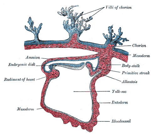
Mesenchyme is characterized morphologically by a prominent ground substance matrix containing a loose aggregate of reticular fibers and unspecialized mesenchymal stem cells. The mesenchyme originates from the mesoderm. This "soup" exists as a combination of the mesenchymal cells plus serous fluid plus the many different tissue proteins. Serous fluid is typically stocked with the many serous elements, such as sodium and chloride. The mesenchyme develops into the tissues of the lymphatic and circulatory systems, as well as the musculoskeletal system.
Mesenchyme , or mesenchymal connective tissue , is a type of undifferentiated connective tissue. It is predominantly derived from the embryonic mesoderm , although may be derived from other germ layers , e. The term mesenchyme is often used to refer to the morphology of embryonic cells that, unlike epithelial cells , can migrate easily. Epithelial cells are polygonal, polarized in an apical-basal orientation, and organized into closely adherent sheets. Mesenchyme is characterized by a matrix that contains a loose aggregate of reticular fibrils and unspecialized cells capable of developing into connective tissue: bone, cartilage , lymphatics and vascular structures. Articles: Intrathoracic sarcoma Retroperitoneal liposarcoma Endometriosis Pseudoangiomatous stromal hyperplasia Fossula post fenestram Facial muscles Soft tissue sarcoma Primary retroperitoneal neoplasms Desmoplastic small round cell tumour of the pleura. Updating… Please wait.
Mesenchyme
Editor's note: Katherine Koczwara created the above image for this article. You can find the full image and all relevant information here. Mesenchyme is a type of animal tissue comprised of loose cells embedded in a mesh of proteins and fluid, called the extracellular matrix.
Cabañas en chihuahua con alberca
Thank you for visiting nature. Normal Table of Xenopus Laevis Daudin. Article Google Scholar. Lee, R. About Recent Edits Go ad-free. Operated neurulae were grown until stage 41—43 1—1. Elsevier Health Sciences. Bodenstein, D. Citation, DOI, disclosures and article data. Development 14, — The focus of mesenchyme research, however, divides between two general interests: the role and expression of mesenchyme-specific genes during development, including pathological processes, and the locations and capabilities of mesenchymal stem cells.
Mesenchyme is a tissue found in organisms during development. It consists of many loosely packed, nonspecialized, mobile cells.
Mapping was according to Bijtel 20 who originally divided the neural plate of stage 16 neurulae along the cranio-caudal axis into 5 rectangular zones. Taken together, these experiments suggest that the posterior neural plate and neural folds region 3 are the main source of tail fin mesenchyme and tail muscle. Epithelial—mesenchymal transition occurs in embryonic cells that require migration through or over tissue, and can be followed with a mesenchymal—epithelial transition to produce secondary epithelial tissues. All animal procedures were performed according to the European Community and local ethics committee guidelines. Article Google Scholar Tucker, A. Center for Biology and Society. Close Please Note: You can also scroll through stacks with your mouse wheel or the keyboard arrow keys. Main article: Epithelial—mesenchymal transition. Figure 6. Experiments on the development of the cranial ganglia and the lateral line sense organs in Amblystoma punctatum. For grafting, vital single cells were randomly picked up with a mouth pipette and transferred homotopically and isochronically one each into white hosts in order to investigate their potency Fig. Using similar strategies in the Mexican axolotl Ambystoma mexicanum and the South African clawed toad Xenopus laevis , we traced the origins of fin mesenchyme and tail muscle in amphibians. D , schematic indicating transverse E—H sectioning planes through ectopic tail. Minot found these cells in the context of histological studies of mesoderm.


I apologise, but, in my opinion, you are mistaken. I can defend the position. Write to me in PM, we will talk.
I am sorry, that has interfered... But this theme is very close to me. Is ready to help.