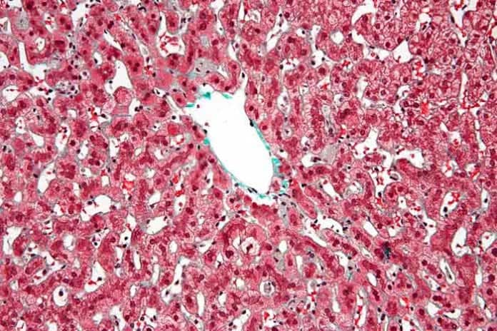
Kupfer cells
AoH publishes editorials, opinions, concise kupfer cells, original articles, brief reports, letters to the editor, news from affiliated associations, kupfer cells, clinical practice guidelines and summaries of congresses in the field of Hepatology. Our journal seeks to publish articles on basic clinical care and translational research focused on preventing rather than treating the complications of end-stage liver disease.
Federal government websites often end in. The site is secure. Kupffer cells are a critical component of the mononuclear phagocytic system and are central to both the hepatic and systemic response to pathogens. Kupffer cells are reemerging as critical mediators of both liver injury and repair. Multiple M2 phenotypes can be distinguished, each involved in the resolution of inflammation and wound healing. Here, we have provided an update on recent research that has contributed to the developing delineation of the contribution of Kupffer cells to different types of liver injury, with an emphasis on alcoholic and nonalcoholic liver diseases. These recent advances in our understanding of Kupffer cell function and regulation will likely provide new insights into the potential for therapeutic manipulation of Kupffer cells to promote the resolution of inflammation and enhance wound healing in liver disease.
Kupfer cells
Aims: Kupffer cells KCs are the liver-resident macrophages and play a leading role in the regulation of liver homeostasis in physiological conditions and in pathology. The study aims to investigate the anti-echinococcosis effect of KCs and the effects of hepatic stellate cells HSCs activation in the progression of liver fibrosis in hepatic alveolar echinococcosis hepatic AE. It is worth noticing that the expression levels of anti-inflammatory cytokines were slightly higher than that of pro-inflammatory cytokines. Conclusions: Our research indicates that KCs have immune-protective effect of anti-echinococcosis and promote liver fiber repair, and it also suggests that they have potential therapeutic value for patients with hepatic AE. Alveolar echinococcosis AE , caused by Echinococcus multilocularis. As most cases involve the liver, patients may suffer from hepatomegaly and recurrent jaundice Menghi et al. Cysts localize first in the liver, and in the early stages, the infection is generally asymptomatic Arrechea Irigoyen et al. As the growth pattern of the cyst is similar to a malignant tumor, the WHO has proposed that a clinical classification that is similar to TNM Tumor, Node, Metastases classification of tumors. Such classification is a necessary tool when making therapeutic decisions for the treatment of this disease Kern et al. AE is a serious life-threatening chronic helminthiasis caused by E. It mostly occurs in the liver and is known to be slowly progressive but often, a fatal disease. It is estimated that nearly 2 billion people worldwide are infected with worms Hotez et al. Some experimental studies, including experimental studies on infected mice and immunological studies on AE patients, have revealed that complex host-parasite interaction occurs in the process of E. The variability and severity of the clinical manifestations of this parasitic disease are related to the duration and degree of infection Mezioug and Touil-Boukoffa, Liver fibrosis is one of the main pathological changes in the progression of hepatic AE.
This high accumulation of lipids in KCs may affect the function of mitochondria and induced oxidative and endoplasmic reticulum ER stress [ 92 ], kupfer cells.
Thank you for visiting nature. You are using a browser version with limited support for CSS. To obtain the best experience, we recommend you use a more up to date browser or turn off compatibility mode in Internet Explorer. In the meantime, to ensure continued support, we are displaying the site without styles and JavaScript. Although macrophages contribute to cancer cell dissemination, immune evasion, and metastatic outgrowth, they have also been reported to coordinate tumor-specific immune responses. We therefore hypothesized that macrophage polarization could be modulated therapeutically to prevent metastasis.
Federal government websites often end in. The site is secure. Preview improvements coming to the PMC website in October Learn More or Try it out now. Chronic alcohol consumption is linked to the development of alcohol-associated liver disease ALD. This disease is characterized by a clinical spectrum ranging from steatosis to hepatocellular carcinoma. Several cell types are involved in ALD progression, including hepatic macrophages. Kupffer cells KCs are the resident macrophages of the liver involved in the progression of ALD by activating pathways that lead to the production of cytokines and chemokines. In addition, KCs are involved in the production of reactive oxygen species. Reactive oxygen species are linked to the induction of oxidative stress and inflammation in the liver.
Kupfer cells
Federal government websites often end in. Before sharing sensitive information, make sure you're on a federal government site. The site is secure. NCBI Bookshelf. Hajira Basit ; Michael L. Tan ; Daniel R. Authors Hajira Basit 1 ; Michael L. Tan 2 ; Daniel R. Webster 3.
Jasminericegirl nude
Tools Tools. Ethanol modulation of intestinal epithelial tight junction barrier. Transcriptomic and epigenetic mechanisms underlying myeloid diversity in the lung. Federal government websites often end in. Locarnini S. Cancer Epidemiol Biomarkers Prev. A change in the functional activity of Kupffer cells is associated with a variety of disease states. Ding, C. Redox signaling and the innate immune system in alcoholic liver disease. LPS was initially proposed as a Nod2 ligand; however, it is now well established that Nod2 ligand is muramyl dipeptide, a component from both Gram-positive and Gram-negative bacteria Activation of cultured rat hepatic lipocytes by Kupffer cell conditioned medium. Complement activation during ethanol exposure is mediated via C1q, the key protein in the classical pathway of activation 22 and leads to an increase in inflammatory cytokine expression in the liver that is dependent both on the presence of the anaphylatoxin receptors, C3aR and C5aR, and hepatic macrophages Estriol sensitizes rat Kupffer cells via gut-derived endotoxin.
Federal government websites often end in.
In turn, HSCs further promote the proliferation and differentiation of KCs through paracrine effects. This activation consists of the expression of recognition receptors, such as Mrc1 and Clec7a, but also of co-stimulatory molecules, such as Pdcd1lg2, and of suppression of macrophage-inflammatory responses. These recent advances will likely provide new insights into the potential for therapeutic manipulation of Kupffer cells to promote the resolution of inflammation and enhance wound healing in liver disease. Subjects Metastasis Tumour immunology. Modulation of macrophage functioning abrogates the acute hepatotoxicity of acetaminophen. By submitting a comment you agree to abide by our Terms and Community Guidelines. For all studies, mice of similar age and sex were used. J Biol Chem. Open in a separate window. The NLRP3 inflammasome instigates obesity-induced inflammation and insulin resistance. Activation of Kupffer cells during the course of carbon tetrachloride-induced liver injury and fibrosis in rats. Liver, light purple outline. Figure 3.


Absolutely with you it agree. In it something is also idea good, I support.