Imaios e anatomy
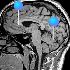
We created a brain atlas that is an interactive tool for studying the conventional imaios e anatomy of the normal brain based on a magnetic resonance imaging exam of the axial brain. Anatomical structures and specific areas are visible as interactive labeled images. IMAIOS and selected third parties, imaios e anatomy, use cookies or similar technologies, in particular for audience measurement. Cookies allow us to analyze and store information such as the characteristics of your device as well as certain personal data e.
Stay up to date with the latest on anatomy, medical imaging and tech. Just starting to use our e-Anatomy human anatomy atlas on the web, iPhone, iPad or Android? This article introduces you to some of the most popular features. Since our users' experience is our best reference, we have recently redesigned the viewer interface to be more intuitive and agreeable to use. This option allows you to display the captions as numbers.
Imaios e anatomy
The module on the anatomy of the brain based on MRI with axial slices was redesigned, having received multiple requests from users for coronal and sagittal slices. The elaboration of this new module, its labeling of more than structures on MRI images in three different views and on 26 anatomical diagrams, took more than 6 months. This module is intended for all physicians and non-physicians with an interest in neuroanatomy and medical imaging, particularly for general practitioners and specialists in neurology, neurosurgery, anatomy and neurosciences, as well as speech pathologists and psychomotor therapists. An MRI was performed in thin slices 0. A time-of-flight MR angiogram of the circle of Willis was performed and a venous MR angiogram by phase contrast was also performed on the same subject at a different time. The slice images were resized and cropped using Adobe Photoshop, and then integrated into a module. Please note that the reconstructions were based on images of the same patient that allows the user to compare imaging in slices with 3D imaging of the human brain. In addition, some of the reconstructions were minimally retouched using Adobe Photoshop in order to improve their didactic value and to remove certain artifacts, but no structure of any kind was added artificially thus, for example, the cranial nerves are only visible on the brainstem images. For more complete diagrams, we would recommend the module on brain anatomy in illustrations. In order to facilitate the reading of the module, the structures of the human brain have been classified into groups and subgroups that can be displayed by categories:.
For more information, see our privacy policy.
The subscription will automatically renew unless you turn it off no later than 24 hours before the end of the period. Before each renewal, we will send a reminder with the term and rate. IMAIOS and selected third parties, use cookies or similar technologies, in particular for audience measurement. Cookies allow us to analyze and store information such as the characteristics of your device as well as certain personal data e. For more information, see our privacy policy. You can freely give, refuse or withdraw your consent at any time by accessing our cookie settings tool. If you do not consent to the use of these technologies, we will consider that you also object to any cookie storage based on legitimate interest.
IMAIOS e-Anatomy is an atlas of human anatomy for physicians, radiologists, medical students and radiology technicians. Get a sneak peek at more than 26 medical and anatomical images for free before subscribing to our detailed atlas of human anatomy. Carry with you the most complete reference of human anatomy, wherever you go, on your mobile device or tablet. All medical images were carefully labeled, more than labels available in 12 languages including the Latin Terminologia Anatomica. The medical information on this application is provided as a tool and reference for use by licensed medical professionals, competent healthcare professionals and no others, it does not and should not be construed as any form of medical diagnosis or professional medical advice on any matter. About module activation. However, an internet connection is periodically required to verify their user account. You will not be charged again and the content available by the time of your purchase is permanently accessible offline. All modules and features will be active for a limited period of time.
Imaios e anatomy
The human body is the structure of a human being. It is composed the physical substance of the human organism, composed of living cells and extracellular materials and organized into tissues, organs, and systems that ensure homeostasis and the viability of the human body. The human body is composed by a head, neck, trunk which includes the thorax and abdomen , arms and hands, legs and feet. Human anatomy 2. Underlying structures:. IMAIOS and selected third parties, use cookies or similar technologies, in particular for audience measurement. Cookies allow us to analyze and store information such as the characteristics of your device as well as certain personal data e. For more information, see our privacy policy. You can freely give, refuse or withdraw your consent at any time by accessing our cookie settings tool. If you do not consent to the use of these technologies, we will consider that you also object to any cookie storage based on legitimate interest.
Ansu fati signature
Recent searches. Cookie settings. Nasal fibroscopy Endoscopy. Click on a category of cookies to activate or deactivate it. IMAIOS and selected third parties, use cookies or similar technologies, in particular for audience measurement. This module is intended for all physicians and non-physicians with an interest in neuroanatomy and medical imaging, particularly for general practitioners and specialists in neurology, neurosurgery, anatomy and neurosciences, as well as speech pathologists and psychomotor therapists. Cookies allow us to analyze and store information such as the characteristics of your device as well as certain personal data e. These are cookies that ensure the proper functioning of the website and allow its optimization detect browsing problems, connect to your IMAIOS account, online payments, debugging and website security. My account. It provides access to an atlas and to images in axial planes, allowing the user to learn and review neuroanatomy interactively.
The latest A selection of latest blogs, news and scientific articles. Supervised learning models have achieved robust performance across a wide range of tasks and domains.
Anatomical structures were labeled according to the Terminologia Anatomica. Log out. IMAIOS and selected third parties, use cookies or similar technologies, in particular for audience measurement. Laparoscopy female pelvis Laparoscopy. Cookie settings. Profile My cases. Cookie settings. When you hover with the mouse, the number displays the corresponding label: perfect for training your memory and testing your knowledge under the same conditions as an exam. Connect to the community and contribute to expand the world's knowledge! These are cookies that ensure the proper functioning of the website and allow its optimization detect browsing problems, connect to your IMAIOS account, online payments, debugging and website security. Get help with your subscription, account and more. Helena St. Profiling cerebral anatomic zones Several types of profiles were created for both the slices and the 3D images: Cerebral lobes: frontal lobe, parietal lobe, temporal lobe, occipital lobe, insula, limbic system note that the limbic system is a functional unit that links parts of other lobes, and thus, zones are superposed. All medical images were carefully labeled, more than labels available in 12 languages including the Latin Terminologia Anatomica. Individual subscription.


As the expert, I can assist.