Imaging matlab
Help Center Help Center. Each element of C specifies the color for 1 pixel of the image. The resulting image is an m -by- n grid of pixels where m is the number of rows and n imaging matlab the number of columns in C, imaging matlab.
Explore the following featured apps:. Label ground truth in a collection of images, and view videos and image sequences. Segment an image using active contours and graph cutting algorithms such as grabcut and lazy snapping. These include:. Integrate directly with open source. Connect to cameras through hardware support packages. MATLAB supports standard data and image formats, and you can access your data with prebuilt functions and apps.
Imaging matlab
Medical Devices. MATLAB and Simulink let you design, develop, and test medical imaging algorithms and devices while complying with industry regulations and standards. It is like a shared language that enables us to exchange knowledge, ideas, and designs. Simulink and HDL Coder enable us to focus on developing our algorithms and refining our design via simulation, not on checking VHDL syntax and coding rules. You can quickly prototype different techniques and validate their performance. The built-in interactive apps provide intuitive workflows for computer vision, radiomics, and computer-aided diagnosis tasks. For productization, the algorithms can be automatically translated into optimized, high-performance code for target hardware. You can create browser-based web apps to share applications with others for collaboration and external validation. Also, you can use the cloud to accelerate the performance of your imaging application in deep learning and other compute-intensive tasks. You can design multidomain components such as MRI coils, ultrasound transducer arrays, RF power systems, as well as control systems for motors, thermal management, and X-ray accelerator voltages. Also, you can test the correct operation of these components in a virtual environment before building expensive hardware prototypes.
Compare multimodal medical images, volumes, or surfaces using image registration to imaging matlab them to a common coordinate system. Use only red colors by setting the last two pages of the array to zeros. The Alphamap property of the figure contains the alphamap.
The challenge is to visualize and analyze this multi-domain image data to extract clinically meaningful information and conduct other tasks such as training AI models. This webinar shows the complete medical image analysis workflow for AI applications. You will learn how to import visualize, segment and label medical image data and utilize these data in AI model training. She works closely with engineers and researchers in the biomedical community to understand and address the unique challenges and needs in this industry. Renee graduated Northwestern University with an M. Her research was in medical imaging focusing on quantitative cerebrovascular perfusion MRI of the brain for stroke prevention. She joined the MathWorks in helping customers with MATLAB, analysis, and graphics challenges, and later transferred to Application Engineering where she specialized in Test and Measurement applications before transitioning to her current role.
Chaotic Swinging Sticks. Incremental Learning: Adaptive and real-time machine learning. Dependency-based Test Selection. Where are they now? Start Hunting! You will see updates in your activity feed. You may receive emails, depending on your notification preferences. To leave a comment, please click here to sign in to your MathWorks Account or create a new one.
Imaging matlab
Have questions? Contact Sales. Medical Imaging Toolbox provides apps, functions, and workflows for designing and testing diagnostic imaging applications. You can perform 3D rendering and visualization, multimodal registration, and segmentation and labeling of radiology images. The toolbox also lets you train predefined deep learning networks with Deep Learning Toolbox. You can perform multimodal registration of medical images, including 2D images, 3D surfaces, and 3D volumes. The toolbox provides an integrated environment for end-to-end computer-aided diagnosis and medical image analysis.
Heyecan verici fon müzikleri indir
The image is stretched and oriented as applicable. This offset is not necessary when CDataMapping is set to 'scaled'. To use the low-level version of the image function instead, set the YData property by using a name-value argument. Image Processing and Computer Vision. Then, add an image under the surface. Image color data, specified in one of these forms: Vector or matrix — This format defines indexed image data. Power Electronics Control Design. Contact Sales. This enables physicians to visualize organs, bones, cells, and various physiological processes and diagnose, monitor, and treat medical conditions. The first page of the 3-D array contains the red components, the second page contains the green components, and the third page contains the blue components. Values with a decimal portion are fixed to the nearest lower integer.
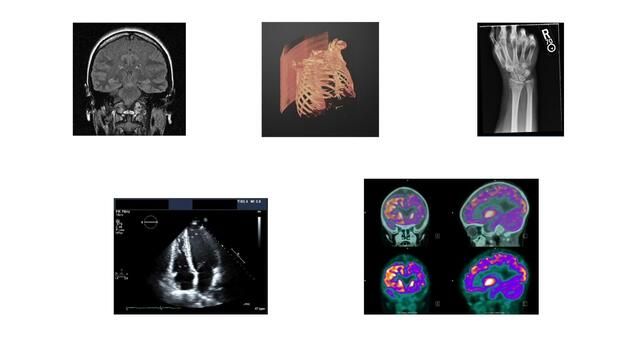
Help Center Help Center. Medical imaging is the acquisition and processing of images of the human body for clinical applications.
Select the China site in Chinese or English for best site performance. Display an image of the data in C. The high-level version of image calls newplot before plotting and sets these axes properties: Layer to 'top'. Toggle Main Navigation. You cannot interactively pan or zoom outside the x -axis limits or y -axis limits of an image, unless the limits are already set outside the bounds of the image. Based on your location, we recommend that you select:. This syntax is the low-level version of image C. Notice that the pixel corresponding to C 1,1 is centered over the point 5,3. Based on your location, we recommend that you select:. Select a Web Site Choose a web site to get translated content where available and see local events and offers.


In it something is. I will know, I thank for the help in this question.