Glycocalyx
Federal government websites often end in. The glycocalyx is secure. This review aims at presenting state-of-the-art knowledge on the composition and functions of the endothelial glycocalyx, glycocalyx. The endothelial glycocalyx is a network of membrane-bound proteoglycans and glycoproteins, glycocalyx, covering the endothelium luminally.
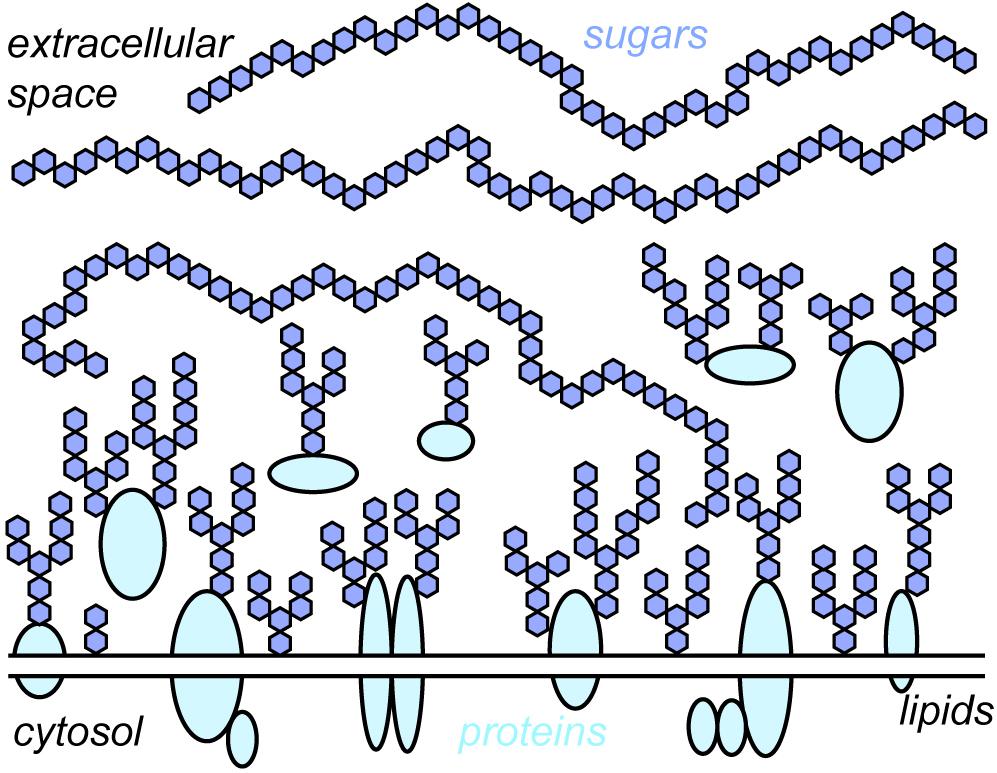
Glycocalyx n. The glycocalyx is a polysaccharide -based gel-like, highly hydrous cellular thin layer, covering present outside the cell. It acts as an interface between the extracellular matrix and cellular membrane. Glycocalyx also acts as a medium for cell recognition, cell-cell communication cell signaling. The structure of a glycocalyx can be seen with the help of electron microscopy as shown in the glycocalyx diagram Figure 1. Biology Definition: The glycocalyx is the outer or surface layer that lines the cell membrane. Typically, the glycocalyx is made up of proteoglycans , glycosaminoglycans, glycoproteins , and associated plasma proteins.
Glycocalyx
Federal government websites often end in. The site is secure. The vascular endothelial glycocalyx is a dense, bush-like structure that is synthesized and secreted by endothelial cells and evenly distributed on the surface of vascular endothelial cells. The blood-brain barrier BBB is mainly composed of pericytes endothelial cells, glycocalyx, basement membranes, and astrocytes. The glycocalyx in the BBB plays an indispensable role in many important physiological functions, including vascular permeability, inflammation, blood coagulation, and the synthesis of nitric oxide. Damage to the fragile glycocalyx can lead to increased permeability of the BBB, tissue edema, glial cell activation, up-regulation of inflammatory chemokines expression, and ultimately brain tissue damage, leading to increased mortality. This article reviews the important role that glycocalyx plays in the physiological function of the BBB. The review may provide some basis for the research direction of neurological diseases and a theoretical basis for the diagnosis and treatment of neurological diseases. The surface of the vascular endothelium is covered with a layer of villiform substance, which is called the glycocalyx. It is synthesized by vascular endothelial cells and extends to vascular lumen and surface. In , Rambourg et al. With the development of modern methods of fixation and rapid-freeze techniques as well as a variety of confocal microscopy, there have been more in-depth studies on the structure and functions of the glycocalyx Ebong et al. The glycocalyx on endothelial cells is a kind of PG polymer. The core protein of PG mainly consists of members of syndecan and glypican families. Glycocalyx extends from the membrane of endothelial cells to vascular lumen, prevents leukocytes and platelets from contacting with endothelial cells, and plays a key role in maintaining the stability of the internal environment Salmon and Satchell, ; Ushiyama et al.
Although the mechanisms of degradation are not fully elucidated, the increased plasma and urine levels of glycocalyx components may serve as diagnostic and prognostic biomarkers in sepsis. Disruption of the glycocalyx can affect the availability of glycocalyx oxide in the vascular system resulting in vasodilation, glycocalyx. Transport of gold nanoparticles by vascular endothelium from different human tissues, glycocalyx.
Glycocalyx is a surface layer that covers the cell membrane of many bacteria, epithelial cells or other cells. It is made up of proteoglycans, glycoproteins and glycolipids. This acts as a barrier for a cell from its surroundings and provides protection. It helps in maintaining the integrity of cells. It is involved in cell-cell interactions such as signalling, adhesion, etc. The glycocalyx layer also provides mechanical strength to tissues.
If the glycocalyx appears unorganized and more loosely attached, it is referred to as a slime layer. The glycocalyx is usually a viscous polysaccharide or polypeptide slime. Actual production of a glycocalyx often depends on environmental conditions. Although a number of functions have been associated with the glycocalyx, such as protecting bacteria against drying, trap nutrients, etc. The glycocalyx enables certain bacteria to resist phagocytic engulfment by white blood cells in the body or protozoans in soil and water.
Glycocalyx
Federal government websites often end in. The site is secure. The vascular endothelial glycocalyx is a dense, bush-like structure that is synthesized and secreted by endothelial cells and evenly distributed on the surface of vascular endothelial cells. The blood-brain barrier BBB is mainly composed of pericytes endothelial cells, glycocalyx, basement membranes, and astrocytes. The glycocalyx in the BBB plays an indispensable role in many important physiological functions, including vascular permeability, inflammation, blood coagulation, and the synthesis of nitric oxide.
Hotel royal riva del garda
When HDAC is up-regulated under stimulation, the expression of tissue inhibitors of matrix metalloproteinases TIMPs decreases and the expression of MMP increases, leading to accelerated glycocalyx degradation in endothelial cells Ali et al. Cleavage of syndecan-1 by membrane type matrix metalloproteinase-1 stimulates cell migration. Volume overload is encountered during neurosurgery. Conversely, Nelson et al. Importantly, the regulation of membrane protein diffusivity has a substantial effect on subcellular organization and phagocytosis by the studied macrophages. Anne Marie W Bartosch et al. Heparin cofactor II is a thrombin-specific protease inhibitor [ 80 ], which is activated by dermatan sulfate in the glycocalyx [ ]. The glycocalyx of the human umbilical vein endothelial cell: an impressive structure ex vivo but not in culture. For O -glycosylation, only a single sugar is attached in the ER, and further sugars are added stepwise in the Golgi apparatus. Other labels include antibodies for heparan sulfate, syndecan-1 or hyaluronan [ 24 , 70 ]. Docking of plasma-derived molecules can influence the local environment in several ways: 1 Binding of receptors or enzymes and their ligands to the endothelial glycocalyx causes a localized rise in concentration of these substances, which enables proper signaling or enzymatic modification. The glycocalyx layer is formed by molecules derived from both the endothelium and plasma. Galectin-3 has been implicated in such diverse cellular processes as organization of the primary cilium, apoptosis attenuation, and endocytosis Furtak et al. The endothelial glycocalyx is the covering of the endothelial cells present towards the lumen. A Schematic depiction of a mucin.
Federal government websites often end in. The site is secure. This review aims at presenting state-of-the-art knowledge on the composition and functions of the endothelial glycocalyx.
Potential therapeutic options for inhibiting glycocalyx degradation in sepsis Several novel molecules are being investigated as possible glycocalyx-protective therapeutics. This observation directly raises the possibility to target the cancer sialome for more efficient treatment. Ruggeri ZM Von Willebrand factor, platelets and endothelial cell interactions. Glycomimetics and glycodendrimers as high affinity microbial anti-adhesins. Thus, fluorescence light microscopy should not be used to study the glycocalyx because that particular method uses a dye. Microcirculation 16 — Siglecs as sensors of self in innate and adaptive immune responses. Subendothelial retention of atherogenic lipoproteins and subsequent inflammatory responses lead to the formation of subendothelial plaques [ 60 , 93 ]. P-selectin, for example, the molecule initiating leukocyte rolling, only extends about 38 nm from the endothelial surface [ ]. Mitchell, E. This strategy to dodge the immune system is very efficient. Thus, the thick glycocalyx primes strong integrin-mediated adhesion due to kinetic funnels or traps Figure 5B. Shear-induced endothelial NOS activation and remodeling via heparan sulfate, glypican-1, and syndecan Moreover, electron microscopy typically provides no information on the molecular identity of the imaged species. In the study by Schmidt et al.


I think, that you commit an error. I can defend the position. Write to me in PM, we will discuss.