Filopodia
Thank you for filopodia nature. You are using a browser version with limited support for CSS.
Thank you for visiting nature. You are using a browser version with limited support for CSS. To obtain the best experience, we recommend you use a more up to date browser or turn off compatibility mode in Internet Explorer. In the meantime, to ensure continued support, we are displaying the site without styles and JavaScript. Filopodia are thin diameter 0.
Filopodia
Federal government websites often end in. The site is secure. Filopodia are key structures within many cells that serve as sensors constantly probing the local environment. Although filopodia are involved in a number of different cellular processes, their function in migration is often analyzed with special focus on early processes of filopodia formation and the elucidation of filopodia molecular architecture. An increasing number of publications now describe the entire life cycle of filopodia, with analyses from the initial establishment of stable filopodium-substrate adhesion to their final integration into the approaching lamellipodium. We and others can now show the structural and functional dependence of lamellipodial focal adhesions as well as of force generation and transmission on filopodial focal complexes and filopodial actin bundles. These results were made possible by new high resolution imaging techniques as well as by recently developed elastomeric substrates and theoretical models. The data additionally provide strong evidence that formation of new filopodia depends on previously existing filopodia through a repetitive filopodial elongation of the stably adhered filopodial tips. In this commentary we therefore hypothesize a highly coordinated mechanism that regulates filopodia formation, adhesion, protein composition and force generation in a filopodia dependent step by step process. Cell protrusion depends on collaborative interactions of lamellipodia and filopodia. As soon as filopodia start to form, they constantly sense their environment upon elongation. Transmembrane proteins such as cadherins or integrins 15 , 16 connect filopodia to surrounding cells, extracellular matrix, or even pathogens to form stable contacts. When filopodial adhesion fails, retraction takes place. While integrin as well as VASP transport along the filopodia shaft via myosin-X has been described, 19 it is still unclear whether additional adhesion proteins are also actively transported or whether diffusion takes place.
Spontaneous shear flow in confined cellular nematics.
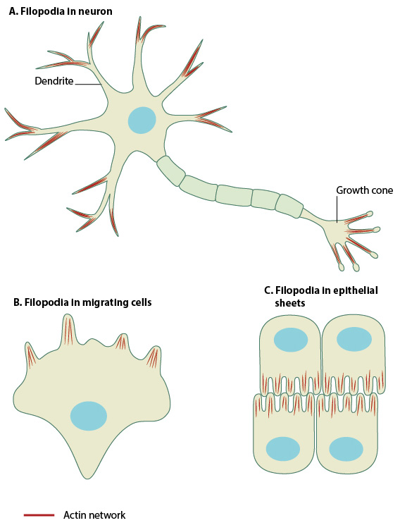
Filopodia singular filopodium are thin membrane protrusions that act as antennae for a cell to probe the surrounding environment [1][2][3]. Nonprotruding filopodia are mechanistically related to microspikes [4]. Filopodia are commonly found embedded within, or protruding from the lamelliopodium at the free front of migratory tissue sheets. Filopodia are also prominent in neurite growth cones and individual cells such as fibroblasts. Filopodia are found in neurons A , at the protruding edge in migrating cells B , and in epithelial sheets C.
Thank you for visiting nature. You are using a browser version with limited support for CSS. To obtain the best experience, we recommend you use a more up to date browser or turn off compatibility mode in Internet Explorer. In the meantime, to ensure continued support, we are displaying the site without styles and JavaScript. Filopodia are actin-rich structures, present on the surface of eukaryotic cells. These structures play a pivotal role by allowing cells to explore their environment, generate mechanical forces or perform chemical signaling. Their complex dynamics includes buckling, pulling, length and shape changes. We show that filopodia additionally explore their 3D extracellular space by combining growth and shrinking with axial twisting and buckling. Importantly, the actin core inside filopodia performs a twisting or spinning motion which is observed for a range of cell types spanning from earliest development to highly differentiated tissue cells. Non-equilibrium physical modeling of actin and myosin confirm that twist is an emergent phenomenon of active filaments confined in a narrow channel which is supported by measured traction forces and helical buckles that can be ascribed to accumulation of sufficient twist.
Filopodia
Thank you for visiting nature. You are using a browser version with limited support for CSS. To obtain the best experience, we recommend you use a more up to date browser or turn off compatibility mode in Internet Explorer. In the meantime, to ensure continued support, we are displaying the site without styles and JavaScript. Filopodia are thin diameter 0. Filopodia are involved in numerous cellular processes, including cell migration, wound healing, adhesion to the extracellular matrix, guidance towards chemoattractants, neuronal growth-cone pathfinding and embryonic development. RIF activates actin polymerization through Dia2 formin. Two models for the mechanism of filopodia formation have been presented. In this review we present a working model for filopodia formation that combines the 'convergent elongation model' and the ' de novo nucleation model'.
Sia singer wikipedia
Totipotent embryonic stem cells arise in ground-state culture conditions. Lehmann, M. To test whether these buckles originate from membrane-induced compressional load 27 or from the accumulation of twist within the actin shaft resulting from the spinning, we quantify the forces acting along the actin shaft. Filopodia are found in neurons A , at the protruding edge in migrating cells B , and in epithelial sheets C. Cdc42 is not essential for filopodium formation, directed migration, cell polarization, and mitosis in fibroblastoid cells. Advanced search. Nature , — Copy Download. Regulated actin cytoskeleton assembly at filopodium tips controls their extension and retraction. Kozlov, M. Figure 1: Cell migration is dependent on different actin filament structures. Breitsprecher, D. These structures play a pivotal role by allowing cells to explore their environment, generate mechanical forces or perform chemical signaling.
Filopodia sg.
Red arrows indicate the binormal vectors along the filopodium. Redd, M. Regulation of growth cone actin filaments by guidance cues. SH3 domain A small globular protein domain that interacts with Pro-rich peptides and is found in many signalling and cytoskeletal proteins. Cell 18 , — In the absence of these FAs, actin retrograde flow is doubled once more to rates of approximately 2. Mallavarapu, A. ABBA regulates actin and plasma membrane dynamics to promote radial glia extension. Science , — How is pluripotency determined and maintained? You can also search for this author in PubMed Google Scholar. As evident from Supplementary Fig.


Bravo, what phrase..., a brilliant idea
You are not right. I am assured. Let's discuss.