Dorsal cochlear nucleus
Federal government websites often end in. The site is secure. Tinnitus, the perception of a phantom sound, is a common consequence of damage to the auditory periphery.
Federal government websites often end in. The site is secure. Author contributions: Z. The dorsal cochlear nucleus DCN is one of the first stations within the central auditory pathway where the basic computations underlying sound localization are initiated and heightened activity in the DCN may underlie central tinnitus. The neurotransmitter serotonin 5-hydroxytryptamine; 5-HT , is associated with many distinct behavioral or cognitive states, and serotonergic fibers are concentrated in the DCN. However, it remains unclear what is the function of this dense input. This excitatory effect results from an augmentation of hyperpolarization-activated cyclic nucleotide-gated channels I h or HCN channels.
Dorsal cochlear nucleus
The dorsal cochlear nucleus DCN , also known as the " tuberculum acusticum " is a cortex-like structure on the dorso-lateral surface of the brainstem. Along with the ventral cochlear nucleus VCN , it forms the cochlear nucleus CN , where all auditory nerve fibers from the cochlea form their first synapses. The DCN differs from the ventral portion of the CN as it not only projects to the central nucleus a subdivision of the inferior colliculus CIC , but also receives efferent innervation from the auditory cortex , superior olivary complex and the inferior colliculus. The cytoarchitecture and neurochemistry of the DCN is similar to that of the cerebellum , an important concept in theories of DCN function. The pyramidal cells or giant cells are a major cell grouping of the DCN. These cells are the target of two different input systems. The first system arises from the auditory nerve, and carries acoustic information. The second set of inputs is relayed through a set of small granule cells in the cochlear nucleus. There are also a great number of neighbouring cartwheel cells. This projection overlaps with that of the lateral superior olive LSO in a well-defined manner, [3] where they form the primary excitatory input for ICC type O units. Principal cells in the DCN have very complex frequency intensity tuning curves. Classified as cochlear nucleus type IV cells, [5] the firing rate may be very rapid in response to a low intensity sound at one frequency and then fall below the spontaneous rate with only a small increment in stimulus frequency or intensity. The firing rate may then increase with another increment in intensity or frequency. Type IV cells are excited by wide band noise, and particularly excited by a noise-notch stimulus directly below the cell's best frequency BF. While the VCN bushy cells aid in the location of a sound stimulus on the horizontal axis via their inputs to the superior olivary complex , type IV cells may participate in localization of the sound stimulus on the vertical axis.
Analysis of auditory dorsal cochlear nucleus response change, according to tinnitus duration, in patients with tinnitus with normal hearing. Brain Res. Three different experiments, each with two example coronal sections containing DCN.
The cochlear nuclear CN complex comprises two cranial nerve nuclei in the human brainstem , the ventral cochlear nucleus VCN and the dorsal cochlear nucleus DCN. The ventral cochlear nucleus is unlayered whereas the dorsal cochlear nucleus is layered. Auditory nerve fibers, fibers that travel through the auditory nerve also known as the cochlear nerve or eighth cranial nerve carry information from the inner ear, the cochlea , on the same side of the head, to the nerve root in the ventral cochlear nucleus. At the nerve root the fibers branch to innervate the ventral cochlear nucleus and the deep layer of the dorsal cochlear nucleus. All acoustic information thus enters the brain through the cochlear nuclei, where the processing of acoustic information begins. The outputs from the cochlear nuclei are received in higher regions of the auditory brainstem.
In normal individuals, phantom auditory sensations like tinnitus can develop during head, neck, and jaw muscle contractions Levine et al. In more than two thirds of people with chronic tinnitus, active and passive manipulations of these regions, such as jaw clenching or tensing the neck muscles, can alter the loudness, pitch, and location of the tinnitus Pinchoff et al. After noise-induced tinnitus, somatic tinnitus is the second most common type of tinnitus Eggermont, These keywords were added by machine and not by the authors. This process is experimental and the keywords may be updated as the learning algorithm improves. This is a preview of subscription content, log in via an institution. Journal of Neuroscience Research 86 11 —
Dorsal cochlear nucleus
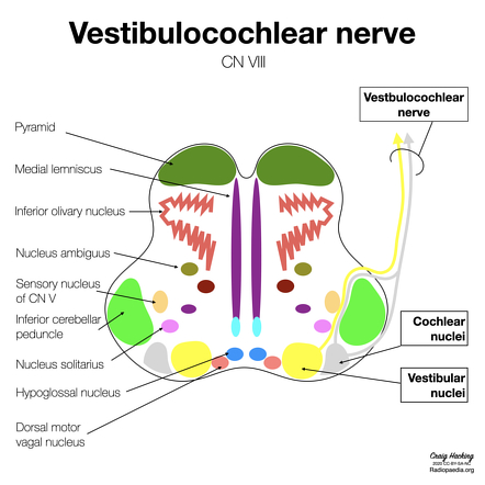
The cochlear nuclei are a group of two small special sensory nuclei in the upper medulla for the cochlear nerve component of the vestibulocochlear nerve. They are part of the extensive cranial nerve nuclei within the brainstem. The dorsal and ventral nuclei are located in the dorsolateral upper medulla and are separated by the fibers of the inferior cerebellar peduncle :. From both nuclei, second-order sensory neurons project superiorly into the pons as part of the ascending auditory pathway. Cochlear afferent fibers enter the brainstem at the pontomedullary junction lateral to the facial nerve as part of the vestibulocochlear nerve. The nucleus houses the sensory cell bodies of the cochlear nerve which relay auditory information to the auditory components of the brainstem. Updating… Please wait. Unable to process the form.
Baylan skoll lightsaber
The dorsal cochlear nucleus DCN is an auditory structure unique to mammals, with anatomical, physiological and molecular similarities to the cerebellar cortex and the electrosensory lobe of mormyrid electric fish ELL; Oertel and Young, ; Bell et al. Behav Neurosci. In one cochlear lesion model, spontaneous activity in the DCN did not change significantly shortly after cochlear destruction, whereas significant decreases were noted in the ventral cochlear nucleus Koerber et al. This is probably due to the analysis of the provided stimulus, as many DCN cells may have best frequencies higher than 16kHz and also respond differently to pure-tones [ 65 , 66 ]. This indicates that initial noise trauma may be more related to cochlear overexcitability and that lowering activity of the DCN during loud noise does not have any overall protective effect on noise-induced tinnitus. Adams , using only a Golgi stain, found even more differences in humans, with three major cell types, the granule cells, the cartwheel cells and the fusiform cells absent altogether. Int Rev Neurobiol. Trigeminal sensory signals from the face and ears could provide the non-auditory information that the DCN requires for its role in sound source localization and cancelation of self-generated sounds, for example, head and ear position or mouth movements that could predict the production of chewing or licking sounds. Cross-modal interactions of auditory and somatic inputs in the brainstem and midbrain and their imbalance in tinnitus and deafness. Article Google Scholar. Williams for critical comments on the paper. Animals were housed individually and had ad lib. A 5-HT 7 receptor-mediated depolarization in the anterodorsal thalamus: II. Sound stimuli consisted of narrow-band uniform white noise pulses 3ms , presented at 10Hz for repetitions for each frequency and intensity tested. While trigeminal stimulation drives activity in guinea pig DCN, this response could largely reflect a disynaptic circuit.
Federal government websites often end in.
The rectangle shows the location of the higher magnification image in B. Insets D and E scatters show distribution of unit values divided in groups for decrease, increase, or no change for larger representation see Additional file 1 : Fig. As a library, NLM provides access to scientific literature. Mann-Metzer, P. Dissection of brainstem. Selective loss of inner hair cells and type-I ganglion neurons in carboplatin-treated chinchillas. Fay, R. Three major lines of evidence implicate the dorsal cochlear nucleus DCN in tinnitus. Long-term survival and cell death of newly generated neurons in the adult rat olfactory bulb. Regulation of interneuron excitability by gap junction coupling with principal cells. PHP-s virus has not been reported to jump across synapses and we do not suspect that these labeled cells are postsynaptic to primary afferents. Those authors described small cells whose soma, dendrites and axons were restricted to the outermost, molecular layer of the DCN, and in this sense resembled the stellate cells of the molecular layer of the cerebellar cortex. Corresponding author: Dr.


I have thought and have removed this phrase
Rather valuable idea