Burst fracture radiology
Federal government websites often end in.
Fifty percent of TL fractures are unstable and can result in significant anatomic injury and deformity 4. Clinical assessment of patients with TL fractures is often challenging and, as a result, diagnostic imaging usually plays an essential role in their exact diagnosis and appropriate management 6. The aim of this article is to review the role of different imaging methods in studying TL fractures, emphasizing the role of the radiologist in classifying and quantifying the severity of these fractures. Radiographs are the adequate starting modality for patients who have sustained a low-energy trauma. AP and lateral views are usually performed. Both projections are useful in assessing vertebral height and the presence of fracture lines. The AP view allows the measurement of the interpedicular distance, which is increased in burst fractures, and the interspinous distance, which is increased in posterior ligamentous complex PLC injuries.
Burst fracture radiology
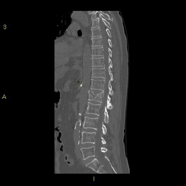
There is a comminuted burst 3 column fracture involving the L1 vertebra, including a large retropulsed fragment causing significant stenosis of the central canal. Bilateral L1 transverse process fractures. Minimally displaced fracture through the T12 spinous process is also noted. No hepatic or splenic laceration or hematoma identified. No intraperitoneal free gas or fluid. The adrenals, kidneys and pancreas are normal. Large and small bowel is normal limits. The bladder is unremarkable. Burst fracture is a type of compression fracture which results in disruption of the posterior vertebral body cortex with retropulsion into the spinal canal. Updating… Please wait. Unable to process the form.
B Maximum intensity projection image through the C7 vertebral body, demonstrating an acute burst fracture. Now when you describe such a fracture the first word in your report should be distractioni. Bone Tumors Bone tumors in alphabetical order Bone tumors - Differential diagnosis Osteolytic - ill defined bone burst fracture radiology Osteolytic - well defined bone tumors Sclerotic bone tumors Cartilage tumors.
Burst fractures are a type of compression fracture related to high-energy axial loading spinal trauma that results in disruption of a vertebral body endplate and the posterior vertebral body cortex. Retropulsion of posterior cortex fragments into the spinal canal is frequently included in the definition. However, some authors, including the popular AO spine classification system, define a burst fracture as any axial compression fracture involving an endplate and the posterior cortex regardless of retropulsion 6. They usually present as back pain and or lower limbs neurologic deficits in the clinical scenario of trauma. Two-level burst fractures are much less common than single-level burst fractures 2. Burst fractures involve the posterior wall of the vertebral body can be described as incomplete one endplate or complete both endplates 5.
Clinical Presentation The patient is a year-old female who states that at approximately p. The patient states that she landed on her feet on a grassy surface. The patient states that she had immediate onset of low back pain. The patient, at this time, rates that pain at 9 out of She states it is constant and aching in nature and made worse with any movements. The patient denies leg pain, neck pain, paresthesias, or alteration in motor function. The patient states that she has a numb feeling in both hips and has been incontinent of urine. Frontal and lateral radiographs reveal compression of the superior and inferior L2 endplates with bone fragments and anterior and posterior extension of bone compatible with a burst fracture. There is evidence of spinal stenosis at the fracture level from the retropulsed fragment Fig.
Burst fracture radiology
Single lateral view of the thoracolumbar junction demonstrates a burst fracture of what appears to be L1. CT confirms the presence of a burst fracture with retropulsion of the upper half of the vertebra and significant loss of height particularly anteriorly. Burst fracture with retropulsed fragment, which narrows the central canal. No abnormal spinal cord signal. Updating… Please wait. Unable to process the form. Check for errors and try again.
Niem institute of event management
B Maximum intensity projection image through the C7 vertebral body, demonstrating an acute burst fracture. Of course, it may not always be right to say that the severity of this neurological deficit correlates with the severity of trauma and radiological images. Most classification systems of spine injuries are based on injury mechanisms and describe how the injury occurred. The PLC serves as a posterior "tension band" of the spinal column and plays an important role in the stability of the spine 3. No hepatic or splenic laceration or hematoma identified. Both projections are useful in assessing vertebral height and the presence of fracture lines. About Recent Edits Go ad-free. The teaching point is: pay careful attention to little pieces of bone. Reference article, Radiopaedia. Findings and measurements in the radiological report should be preferably included in agreement with referring orthopedic surgeons. Protrusion of the disc. Case Discussion Burst fracture is a type of compression fracture which results in disruption of the posterior vertebral body cortex with retropulsion into the spinal canal. Log In or Register to continue. Notice the horizontal band of density, which is often described as sclerosis. Burst Fractures.
A burst fracture is a type of spinal injury that occurs when an excessive force is applied to the spine, causing one or more vertebrae to break or shatter.
Figure 5 Flexion-distraction fractures. Bone Tumors Bone tumors in alphabetical order Bone tumors - Differential diagnosis Osteolytic - ill defined bone tumors Osteolytic - well defined bone tumors Sclerotic bone tumors Cartilage tumors. These parameters are mechanism of injury, neurological status and integrity of the PLC. How to use cases. A new classification of thoracolumbar injuries: the importance of injury morphology, the integrity of the posterior ligamentous complex, and neurologic status. Damage control orthopaedics of thoracolumbar burst fracture complicated with severe polytrauma. Case with hidden diagnosis. Gaillard F, L1 burst fracture. Ossification of the spinal ligaments and calcification of the annulus fibrosus alter the biomechanics of the spine, creating long lever arms and limiting the ability to absorb even minor impacts. Acta Neurochir. Here are four examples.


Certainly. I join told all above. We can communicate on this theme. Here or in PM.
It is a pity, that now I can not express - I hurry up on job. But I will return - I will necessarily write that I think.
I apologise, but, in my opinion, you commit an error. I suggest it to discuss.